
Figure 4.
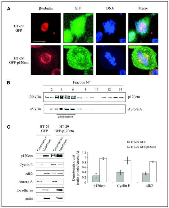
p120ctn localizes to the mitotic furrow and the centrosomes in mitotic HT-29 cells. A, HT-29 GFP (top) and HT-29 GFP-p120ctn (bottom) cells were grown for 48 h on coverslips, fixed with 3% paraformaldehyde, and immunostained for β-tubulin before observation under a confocal microscope DNA was stained blue with 4′,6-diamidino-2-phenylindole (DAPI). Chosen fields are representative of mitotic cells in each condition. Bar, 10 μm. B, centrosome fractions were enriched from mitotic HT-29 GFP-p120ctn cells using a discontinuous sucrose gradient (see Material and Methods). Antibodies against p120ctn and Aurora A were used for Western blotting analyses of each collected fraction. C, left, centrosome- and cell membrane–enriched fractions obtained from the previously described discontinuous sucrose gradient were analyzed by Western blotting using antibodies against p120ctn, cyclin E, cdk2, Aurora A, E-cadherin, and actin. Right, histograms represent the densitometric ratio of p120ctn, cyclin E, and cdk2 to Aurora A levels in centrosomal fractions. Columns, mean of three independent experiments; bars, SE.