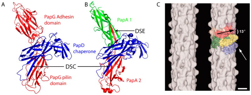
Fig. 2. Donor strand complementation (DSC) and donor strand exchange (DSE) as observed in crystal structures and cryo-EM map of the P pilus rod.

(A) Crystal structure of the PapD-PapG chaperone-adhesin complex (PDB ID: 1QUN). (B) Crystal structure of a PapD-PapA1-PapA2 complex (PDB ID: 2UY6). The arrows labeled DSC indicate the chaperone G1 β-strand engaged in DSC with the pilin domain of PapG (A) or the PapA2 subunit (B). The arrow labeled DSE indicates the Nte of the PapA2 subunit engaged in DSE with the PapA1 subunit (B). (C) Cryo-EM map (left) and modeling with the crystal structure of PapK (right) of the P pilus rod at an estimated resolution of 10 Å. The major pilin PapA appears to sit 13° tilted from horizontal in the helical rod. The white arrow points to a surface protrusion that is presumably the hinge loop formed by the PapA N terminus. Scale bar: 25 Å. Panel C is adapted from reference [25].
