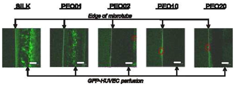
Figure 8.

Perfusion of GFP-transduced human umbilical vein endothelial cells (HUVECs). Silk microtubes embedded in a collagen gel were perfused with green fluorescent protein (GFP) transduced HUVECs. GFP-HUVECs were perfused back and forth at a rate of 4 μL/min through the silk microtubes using a syringe pump, and fluorescent confocal microscopy images are shown after 3 days of perfusion. Sections of the microtubes are shown at the right of each image with GFP-HUVECs present in the channel of the microtube (edge of microtube given by dashed line). These cells are visible in the 100% silk fibroin and PEO01 tubes, but their presence in the microtubes with greater percentages of PEO is obscured due to the opaqueness of the tubes. The silk microtubes were a significant barrier to cell migration. At low porosity, GFP-HUVEC migration was completely blocked; at higher porosities (PEO10 and PEO20) cell migration was limited to a few cells (outlined by red-dashed ovals) over the entire microtube. Scale bars = 300 microns.
