
Figure 6.
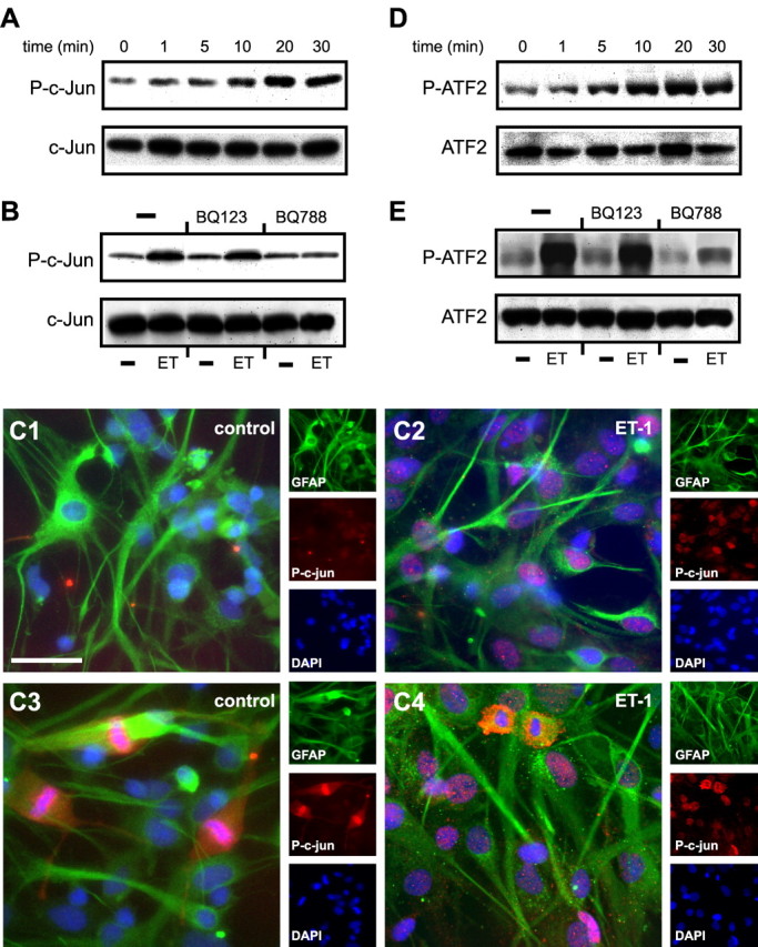
ET-1 induces c-jun and ATF2 phosphorylation in astrocytes. A, D, Time course of ET-1-induced c-Jun (A) and ATF2 (D) phosphorylation. Cells were stimulated with 100 nm ET-1 for the indicated times, and total cell lysates were analyzed by Western blot using an anti-P-c-Jun antibody or an anti-P-ATF2 antibody. The same blots were then stripped and incubated with phosphorylation-state independent anti-c-Jun or anti-ATF2 antibodies to normalize for the total (phosphorylated plus nonphosphorylated) amounts of c-Jun (A, bottom) and ATF2 (D, bottom). C, P-c-Jun immunostaining of cultured astrocytes incubated with saline (C1, C3) or ET-1 (200 nm; C2, C4) for 20 min. In quiescent, unstimulated astrocytes, P-c-Jun was only detected in GFAP+ cells in metaphase (C3), whereas the majority of ET-1-treated astrocytes expressed high levels of nuclear P-c-Jun (C2, C4). Small panels show immunostaining for individual antigens in the same microscopic field. Scale bar, 50 μm. B, E, ET-1-induced c-Jun and ATF2 phosphorylation is selectively blocked by the ETB-R antagonist BQ788, but is not modified by the ETA-R antagonist BQ123. The same blots were then stripped and incubated with phosphorylation-state independent anti-c-jun or anti-ATF2 antibodies to normalize for the total (phosphorylated plus nonphosphorylated) amounts of c-jun (B, bottom) and ATF2 (E, bottom). Data are representative of three independent experiments.