
Figure 7.
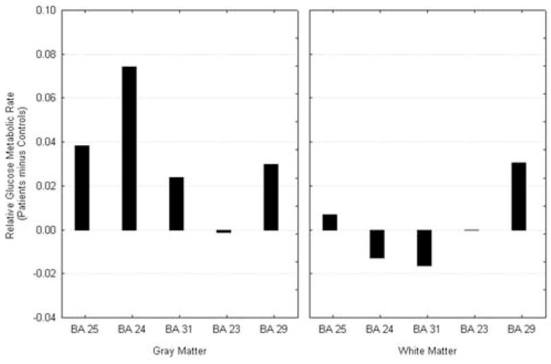
Cingulate Activity at Saline condition. Cingulate gyrus arch shows patients with relatively higher activity than normals in the anterior portion and lower in the midsection; white matter shows decreases throughout.
Official websites use .gov
A
.gov website belongs to an official
government organization in the United States.
Secure .gov websites use HTTPS
A lock (
) or https:// means you've safely
connected to the .gov website. Share sensitive
information only on official, secure websites.
Cingulate Activity at Saline condition. Cingulate gyrus arch shows patients with relatively higher activity than normals in the anterior portion and lower in the midsection; white matter shows decreases throughout.