
Figure 1.
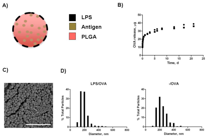
Characterization of nanoparticles. A) Schematic of biodegradable particles with lipopolysaccharide modification. B) Controlled release profile of OVA from LPS/OVA (●) and -/OVA (■) particles. Figure represents release from nanoparticles at 37°C in phosphate-buffered saline in triplicate over 3 weeks. C) Scanning Electron Microscopy (SEM) image of LPS-modified nanoparticles. Scale bar is 2 μm. D) Size distribution profiles for LPS-modified and unmodified OVA-loaded nanoparticles.