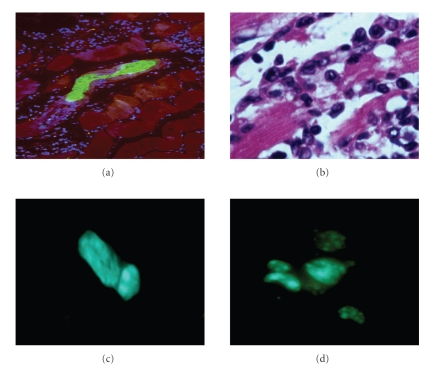
Figure 1.
Heart sections of T. cruzi-infected mice. (a) BALB/c mouse during the acute phase of infection with Colombian strain T. cruzi, showing a parasite nest (green), DAPI-stained nuclei (blue) and myofibers (red). The majority of the DAPI nuclei belong to inflammatory cells that infiltrate the heart in areas infected with parasites. (b) Inflammation of chronic chagasic BALB/c mouse, showing inflammatory cells adhered to myofibers causing myocytolysis. (c) and (d), Detection of bone marrow stem cells (BMC) in the myocardium of chronic chagasic mice. BMC obtained from normal BALB/c mice were injected i.v. into chronic chagasic mice (18 months of infection). BMC were incubated with the fluorescent DNA stain Hoechst 33258 prior to injection into chagasic mice. Sections of frozen heart fragments were prepared 7 (c) and 15 (d) days after BMC injection and fixed with cold acetone. Sections were observed in an Olympus spectral confocal microscope FV1000 observed by fluorescence microscopy. In chagasic mice Hoechst+ cells could be observed 1–7 days after BMC injection, some of which were already beginning cell division cycles (c). Hoechst+ cells proliferated and formed clusters of cells bearing a dotted nuclear fluorescent pattern that could be observed up to 30 days after BMC transplant (d).

