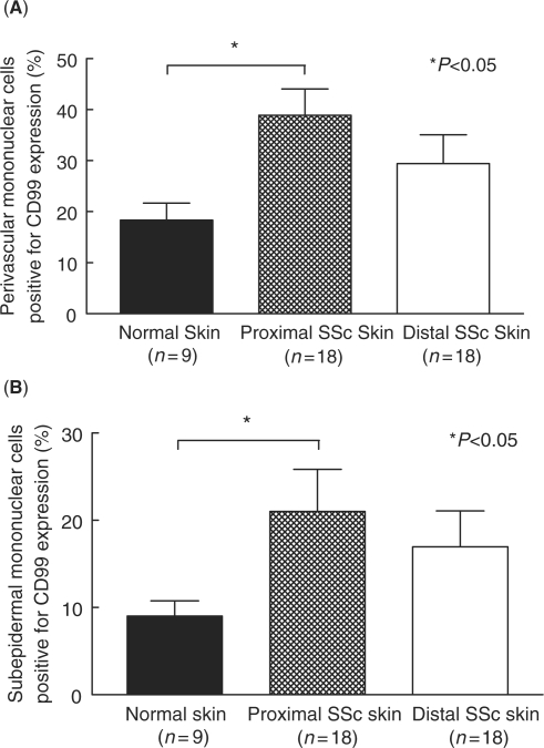
Fig. 2.
Immunohistochemical analysis of CD99 on SSc and normal skin mononuclear cells. Frozen sections of proximal and distal SSc and normal skin were immunostained for CD99. (A) CD99 expression was increased on perivascular mononuclear cells in proximal (39%) and distal (29%) SSc skin compared with normal skin (18%). (B) CD99 expression was increased on subepidermal mononuclear cells in proximal (21%) and distal (17%) SSc skin compared with normal skin (9%). Means are given with the SEM. n = number of patients. P< 0.05 was considered significant.

