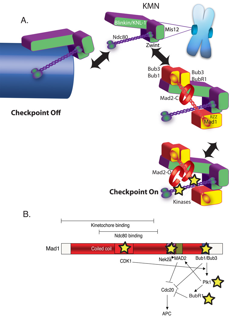
Figure 2. An updated model for spindle checkpoint signaling.
A) Dual role of KMN in the kinetochore as both microtubule anchor and a scaffold to generate spindle checkpoint signals. Overall color scheme is the same as Figure 1. KMN (purple and green) is located in the outer plate of the kinetochore where both binds microtubules (arrow left) and spindle checkpoint protein (arrow right). KMN contains at least two microtubule-binding interfaces one in the KNL-1 subunit and another in the Hec1 subunit of the Ndc80 complex and approximately eight KMNs generate a binding pocket (not shown). In the absence of microtubules KMN has both direct and indirect interactions with checkpoint proteins. KNL-1 binds Bub3/Bub1 and Bub3/BubR1. The Ndc80 subunit can bind a coiled-coil region of Mad1 in a two-hybrid assay. Finally through the Zwint protein, the Mis12 complex binds RZZ, which can strip Mad1 from kinetochores. The kinetochore activates the checkpoint by acting as a scaffold to recruit and activate Bub1 and BubR1 kinases as well as other kinases recently implicated in checkpoint signaling (stars). B) A schematic Map of the Mad1 protein highlighting kinase interactions (stars) and a potential signal transduction network initiated after Mad1 recruitment to the kinetochore.

