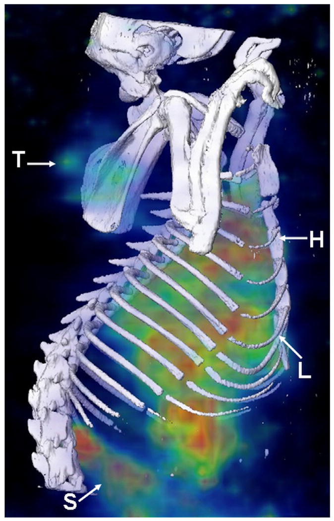
Figure 3.

MicroSPECT/CT images acquired at 20 h post administration of 186Re-Doxil using MPH collimator. Three dimensional (3D) volume rendered SPECT image of 186Re-Doxil overlaid with CT isosurface displayed in bone window shows the accumulation in Tumor (T), liver (L), spleen (S) and circulation through heart (H).
