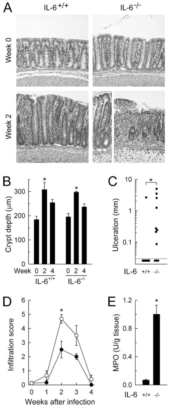
FIGURE 4.

Histological analysis of infection-associated colitis in IL-6-deficient mice. IL-6-deficient mice (IL-6−/−, ○) and wild-type controls (IL-6+/+, ●) were infected orally with C. rodentium or left uninfected as controls (wk 0). The colon was removed at the indicated times, paraffin sections were prepared, stained with H&E, and assessed for damage and inflammatory cell infiltration. A, Representative colon areas, showing similar hyperplasia but greater epithelial ulceration and mucosal infiltration with inflammatory cells after infection in IL-6-deficient mice compared with wild-type controls. Crypt depths (B) and epithelial ulceration (C) were determined morphometrically, and infiltration of mucosa and submucosa with inflammatory cells was scored semiquantitatively (D). Colon homogenates were analyzed for neutrophil infiltration by enzymatic assay for myeloperoxidase (MPO) activity (E). Uninfected mice had no detectable myeloperoxidase activity in the colon (<0.01 U/g tissue). Data are mean ± SEM of three or more mice (B, D, and E) or are results from individual mice (C). *, p < 0.05.
