
FIGURE 6.
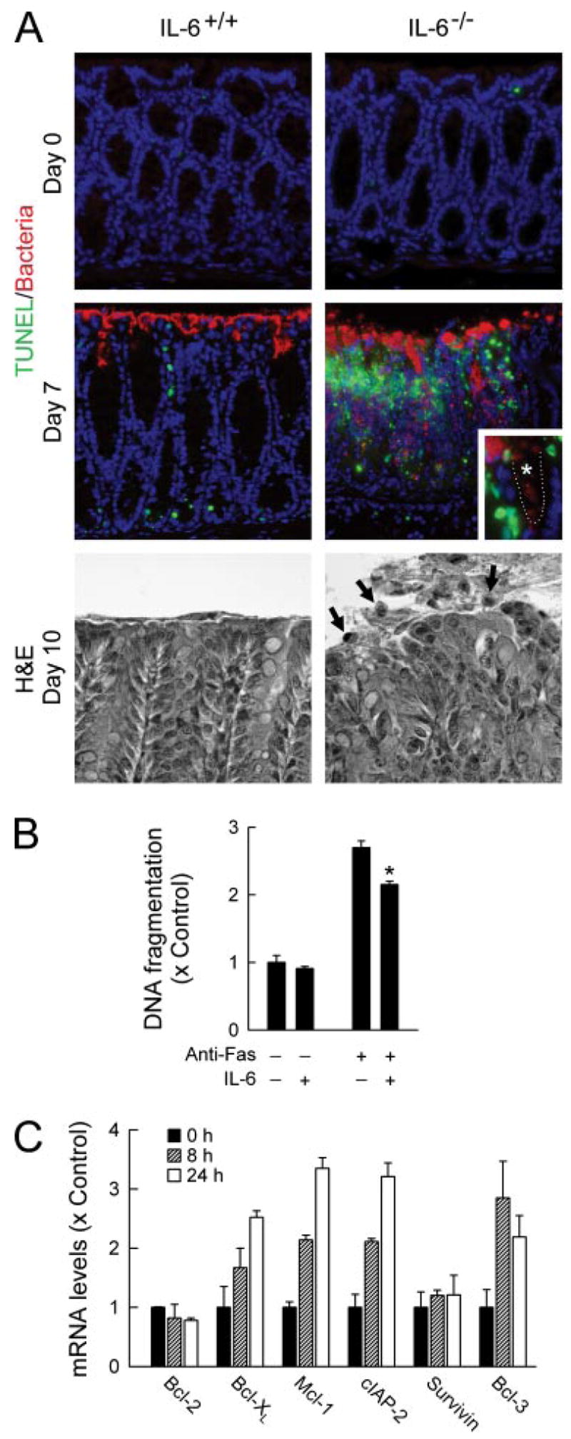
IL-6-dependent protection of colon epithelial cells against apoptosis. A, IL-6-deficient mice (IL-6−/−) and wild-type controls (IL-6+/+) were infected orally with C. rodentium or left uninfected as controls (day 0). The colon was removed at the indicated times, paraffin sections were prepared, double-stained for apoptotic cells by the TUNEL technique (green) and for C. rodentium by specific Abs (red), and counterstained with 4′,6′-diamidino-2-phenylindole (A, top). Inset, crypt region of an IL-6-deficient mouse at higher magnification. ....., Epithelial surface; *, crypt lumen. Parallel sections were stained with H&E (A, bottom). Arrows, apoptotic cells. B, T84 colon epithelial cells were incubated with or without IL-6 for 24 h and treated with anti-Fas Abs (clone CH-11) for an additional 18 h. Apoptosis was assayed with a nucleosome release ELISA. Values are mean ± SD (n = 3) and are expressed relative to unstimulated controls. *, p < 0.05 compared with cells not treated with IL-6. C, T84 cells were stimulated with 100 ng/ml IL-6 for 8 or 24 h or were left unstimulated (0 h), and mRNA levels for the indicated genes were determined by real-time RT-PCR. Values are mean ± SD (n = 3) and are expressed relative to unstimulated controls.