
FIGURE 8.
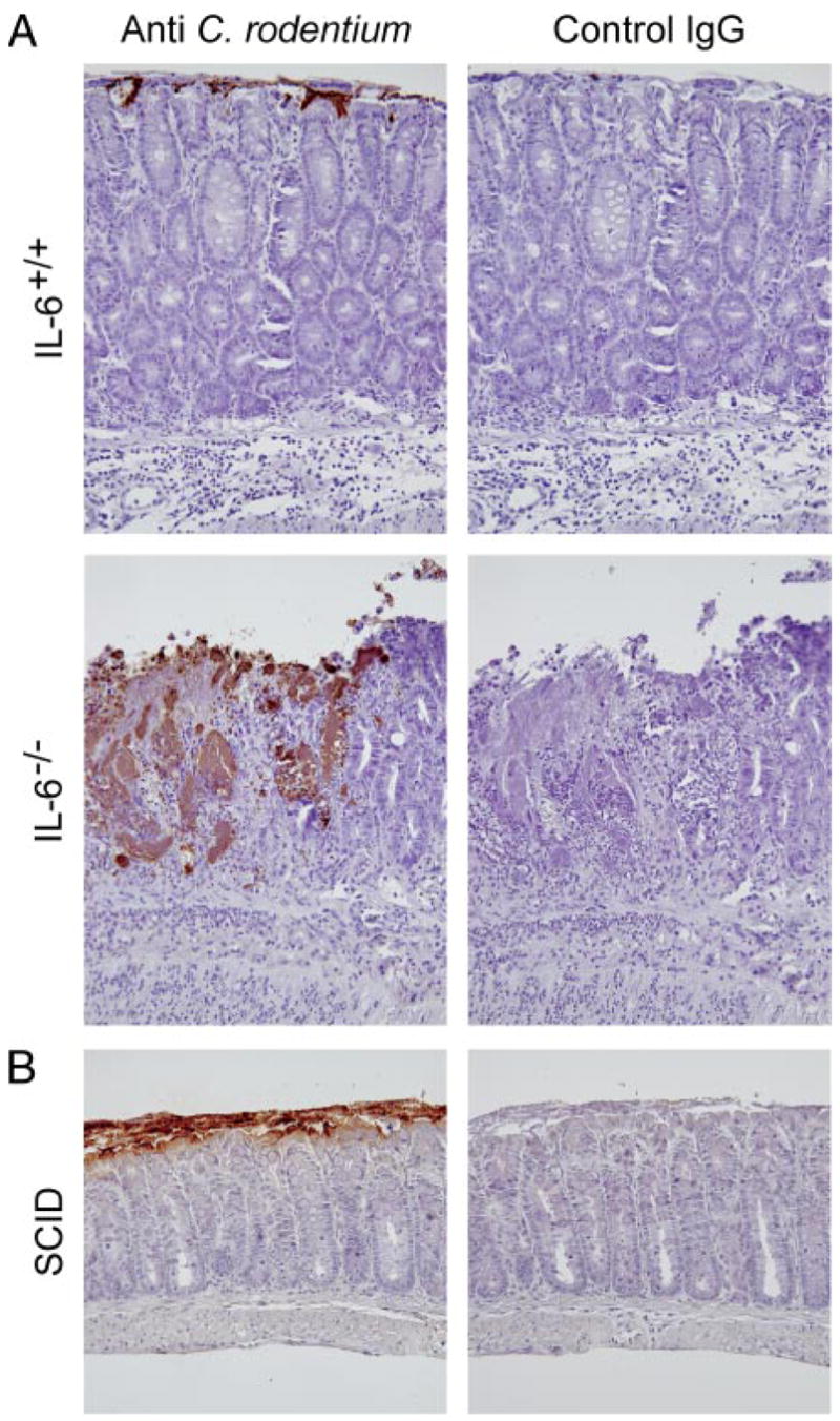
Bacterial localization in the colon of IL-6-deficient mice. Colon segments from IL-6-deficient (IL-6−/−, A) and wild-type mice (IL-6+/+, A) or SCID mice (B) were collected 2 wk after C. rodentium infection, and paraffin sections were stained by an indirect immunoperoxidase method with Abs against C. rodentium or with control IgG Abs. No specific staining was observed in uninfected mice in any of the groups.