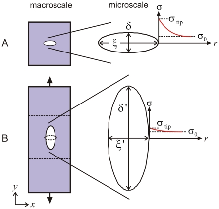
Figure 8. Change of the microscopic crack shape as the protein network undergoes macroscopic mode I tensile deformation.
Panel A shows shape of the initial crack (an elliptical geometry where the length in the x-direction is much greater than the extension in the y-direction). Panel B shows shape of the final crack before onset of failure, representing an elliptical geometry where the length in the y-direction is much greater than the extension in the x-direction. The plots also indicate the distribution of stresses for both cases (the solution for the stress field is symmetric, but shown here only for the right half). The crack shapes reflect those measured in the simulations shown in Figure 6 (there highlighted in white color). The initial geometry and crack shape is shown in panel B (left part) in dashed lines to illustrate the significant transformation.

