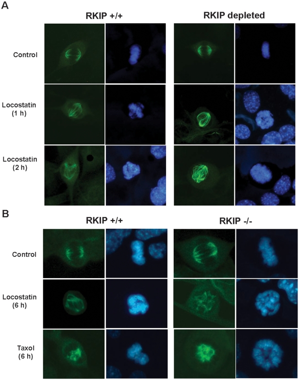
Figure 7. Locostatin disrupts the mitotic spindle and chromosome organization.
MEFs expressing (A) wild-type (RKIP+/+) or lacking RKIP were plated at 2×10⁴ cells/coverslip and grown for 48 h before treatment with either DMSO (Control) or locostatin (20 µM) for either 1 or 2 h. (B) Wild-type (RKIP+/+) or deficient (RKIP−/−) RKIP were plated as described in (A) and treated with either DMSO (Control), locostatin (20 µM) or paclitaxel (10 µM) for 6 h. Cells were fixed and permeabilized and stained with anti-α-tubulin antibody-green. DNA/chromatin was stained with Höechst 33342-blue. Digital photomicrographs were captured using Openlab Darkroom (Improvision) and a Retiga 1300 color digital camera (Q Imaging). Intensity of images were adjusted to display qualitative rather than quantitative comparisons so that the intensity of staining in each panel cannot be directly compared.

