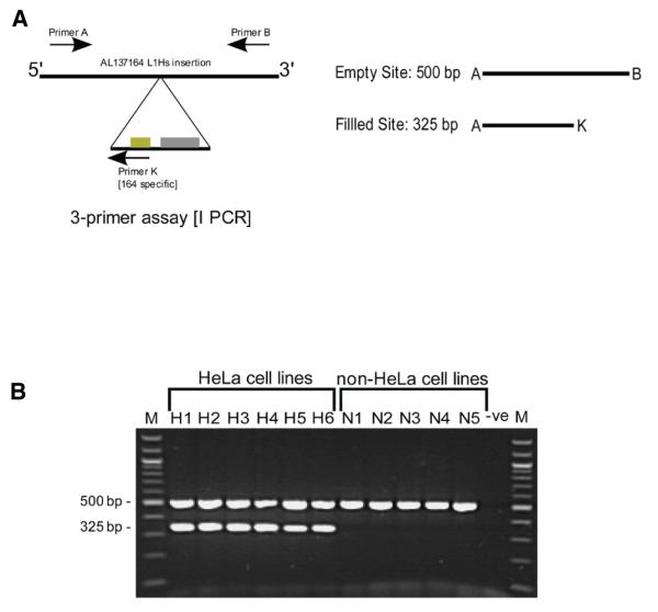
Figure 2. Duplex PCR design and genotyping results.
(A) A duplex PCR assay for insertion AL137164. Primers A and B, specific to the AL137164 insertion site, were combined with a primer (K) specific to the junction between the L1 insertion and its 5′ flanking genomic DNA. The primers generate two amplicons of 500 bp and 325 bp from the empty and filled sites, respectively. (B) PCR amplification using the three-primer duplex assay. Genomic DNA from 6 independently sourced HeLa cell lines (H1, H2, H3, H4, H5, and H6) and 5 different non-HeLa cell lines (N1, N2, N3, N4, and N5) was subjected to PCR amplification. M, 100-bp ladder molecular weight marker; Lanes H1–H6, three-primer amplification of HeLa genomic DNA; Lanes N1–N5, three-primer amplification of non-HeLa cell line genomic DNA; -ve, negative control reaction (no input DNA).

