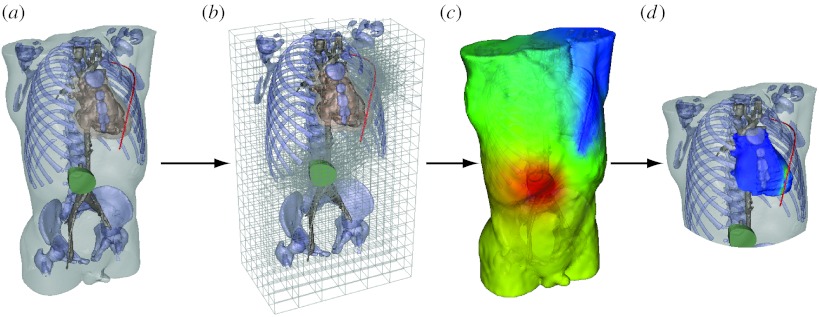
Figure 6.
Pipeline for computing defibrillation potentials in children. The figures shows the steps ((a) setting electrode configuration, (b) refinement of hexahedral mesh for electrode locations, (c) finite-element solution of potentials and (d) analysis of potentials at the heart to predict defibrillation effectiveness) required to place electrodes and then compute and visualize the resulting cardiac potentials.

