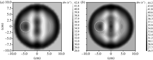
Figure 11.
Reconstructed MWT images of a simulated brain model with a stroke injury with radius 2 cm located at {−4, 0} obtained using multi-frequency reconstruction: (a) 0.5 and 1.0 GHz; and (b) 2.0 and 1.0 GHz. 1% noise. Area with suspected stroke injury is circled in white. Reconstruction using a three-dimensional gradient method.

