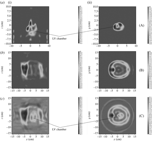
Figure 7.
Reconstructed MWT images of (a) excised swine heart, (b) swine torso and (c) swine torso with heart for ϵ′. (i) Longitudinal view (Y=0) and (ii) transverse view (Z=0). Frequency 0.9 GHz. Scales are in cm. (Note the differences in scales between case (a) and cases (b) and (c).) LV, left ventricular chamber. Reconstruction using a three-dimensional gradient method.

