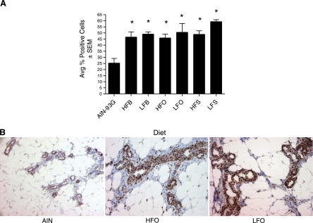
Fig. 2.
PCNA immunohistochemistry of mammary ducts and lobules for pups on different fatty acid diets at postnatal day 50 (PND50). Mammary ducts and lobules stained with PCNA (Leica Clone PC10) and biotinylated horse anti-mouse IgG (Vector) were photographed at ×20 with a Zeiss Axioscope (Carl Zeiss MicroImaging) from the reference diet and low fat (10% of energy as fat) and high fat (39% of energy as fat) olive, safflower, and butter diets. A: for each animal, at least 100 cells from 10 lobular areas were counted with respect to PCNA positivity and negativity, which is expressed as percent, and animals within treatment groups were averaged and compared with AIN-93G. B: examples of PCNA staining in offspring on AIN-93G and HFO and LFO diets for PND50. The percent of PCNA-positive cells in these 3 particular lobular areas were 14.4, 42.7, and 48.2% for AIN-93G HFO and LFO diets, respectively.

