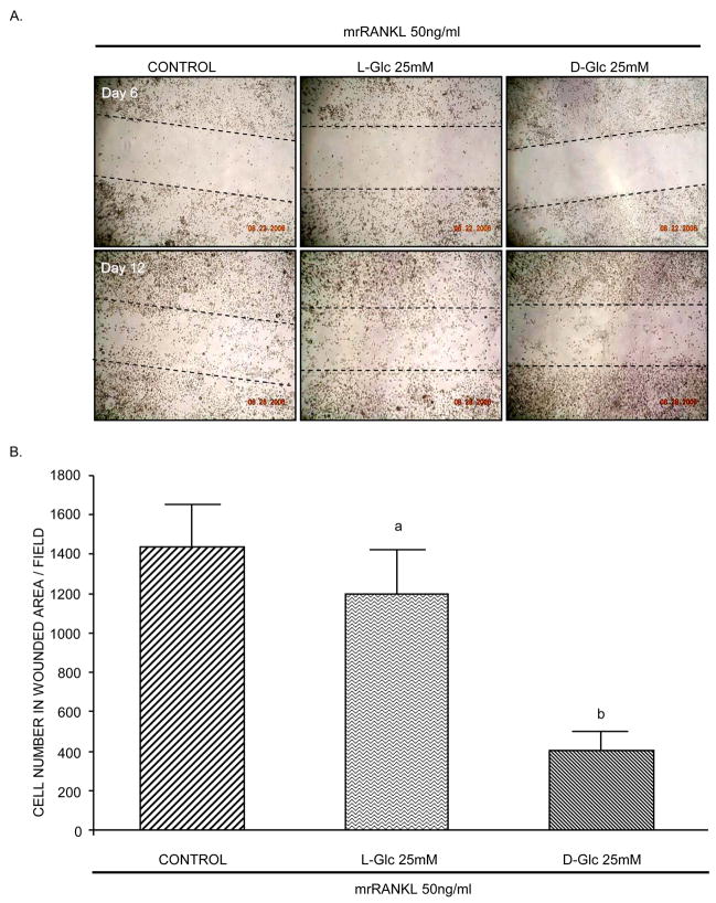
Figure 5. Effect of glucose on cell migration.
A) RAW264.7 cells were seeded at a density of 1×10⁴ cells/cm2 and incubated with mrRANKL (50ng/ml) for 12 days in the presence or absence of high L-Glc or high D-Glc. The medium was changed every 2 days. On day 6, cell monolayers were wounded, washed three times with serum-free α-MEM medium and continued for 6 additional days. Photomicrographs are representative of three independent experiments. B) Cells present in wounded area were counted on day 12. Values are means +/− S.E. of three independent experiments. a (p)>0.05 vs control; b (p)<0.001 vs control or L-Glc-treated cells.

