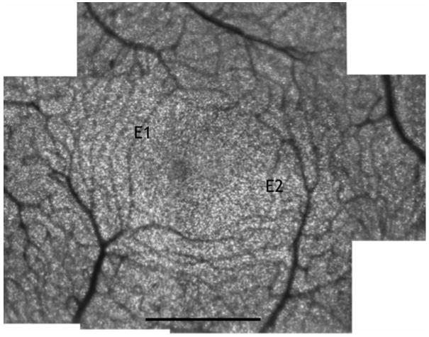
Figure 1.
Foveal montage of subject E. The image is comprised of a series of registered images from AOSLO videos. The two capillaries used for leukocyte velocity and pulsatility calculations are marked E1 and E2. The original data was sampled at 205 pixels per degree (1.43 micrometers per pixel for this subject) Scale bar = 500μm.

