Fig. 3.


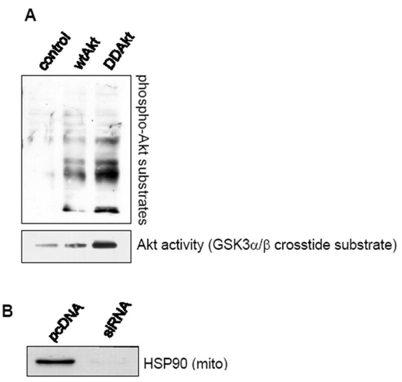
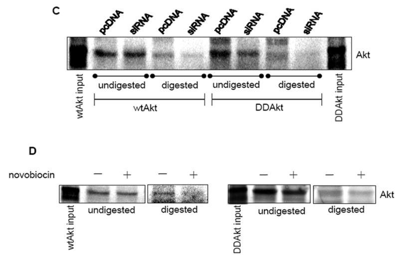
HSP90 mediates the mitochondrial translocation of Akt. (A) Top panel: Isolated mitochondria were incubated with either wild type (wt) or DDAkt, digested with proteinase K, and immunoblotted with a phospho-Akt substrate antibody. Bottom panel: In-vitro translated Akt proteins were measured for enzymatic activity as described in Material and methods. (B) Mitochondria were isolated from pcDNA control cells and HSP90 siRNA cells. Mitochondrial lysates were immunoblotted for HSP90. (C) Representative autoradiograph showing mitochondrial translocation of 35S-Methionine labeled wtAkt and DDAkt in control cells and HSP90 siRNA cells. Isolated mitochondria from each cell line were incubated with radiolabled exogenous wtAkt or DD-Akt protein. Half of each sample was digested with proteinase K, and the other half was left undigested. The Akt bands were viewed with a phosphorimager. (D) Representative autoradiograph showing mitochondrial translocation of wtAkt (left panel) and DDAkt (right panel) in the presence and absence of 625 μM novobiocin.
