Fig. 5.


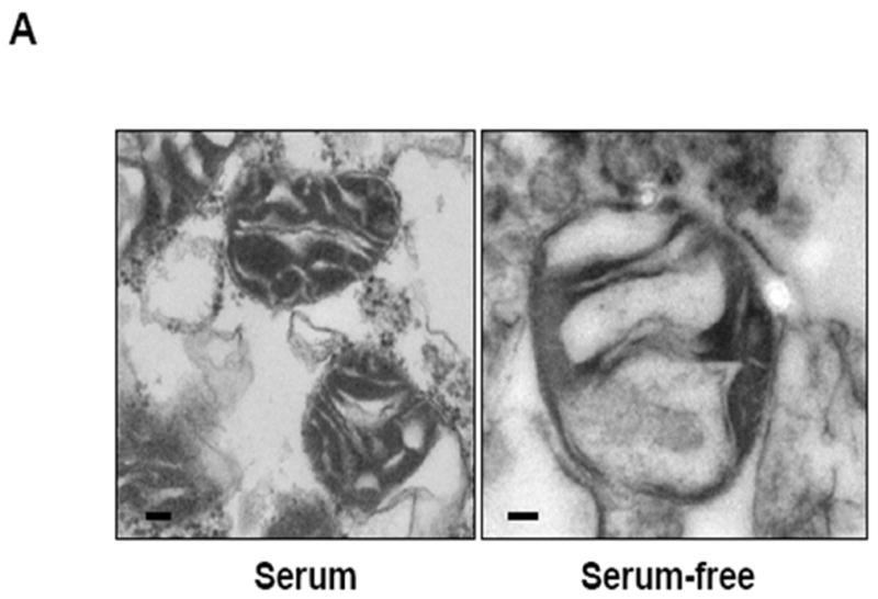
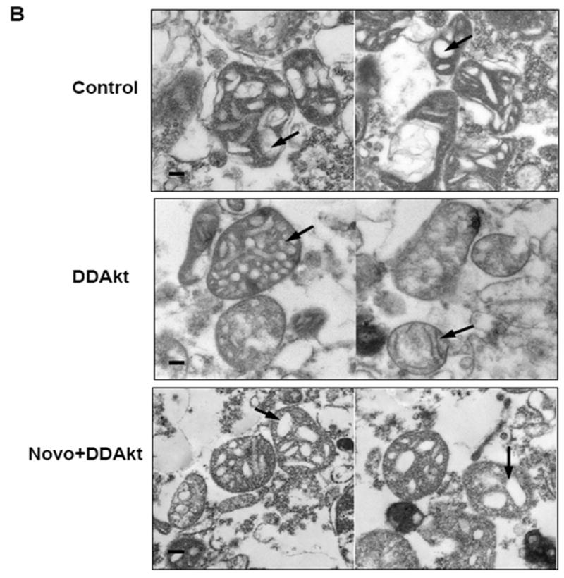
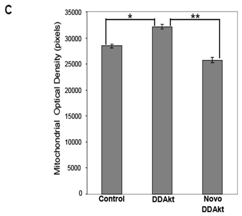
Akt affects mitochondrial morphology. (A) Mitochondrial pellets were fixed and prepared for transmission electron microscopy as described in the Materials and Methods section. Representative transmission electron micrographs of mitochondria isolated from HEK293 cells in serum-containing versus serum-containing media (size bar = 100 nm). (B) Mitochondria were isolated from serum-starved HEK293 cells and incubated with empty reticulocyte (control), DD-Akt, or 625 μM NB and DD-Akt for 45 minutes (size bar = 100 nm). Representative images are shown. (C) Optical density was quantified in a blind study from transmission electron micrographs of isolated mitochondria incubated with DD-Akt or NB + DD-Akt versus control. N=54, *p<0.05, compared to control mitochondria incubated with reticulocyte lysate. **p<0.05, compared to control mitochondria incubated with DD-Akt. ANOVA.
