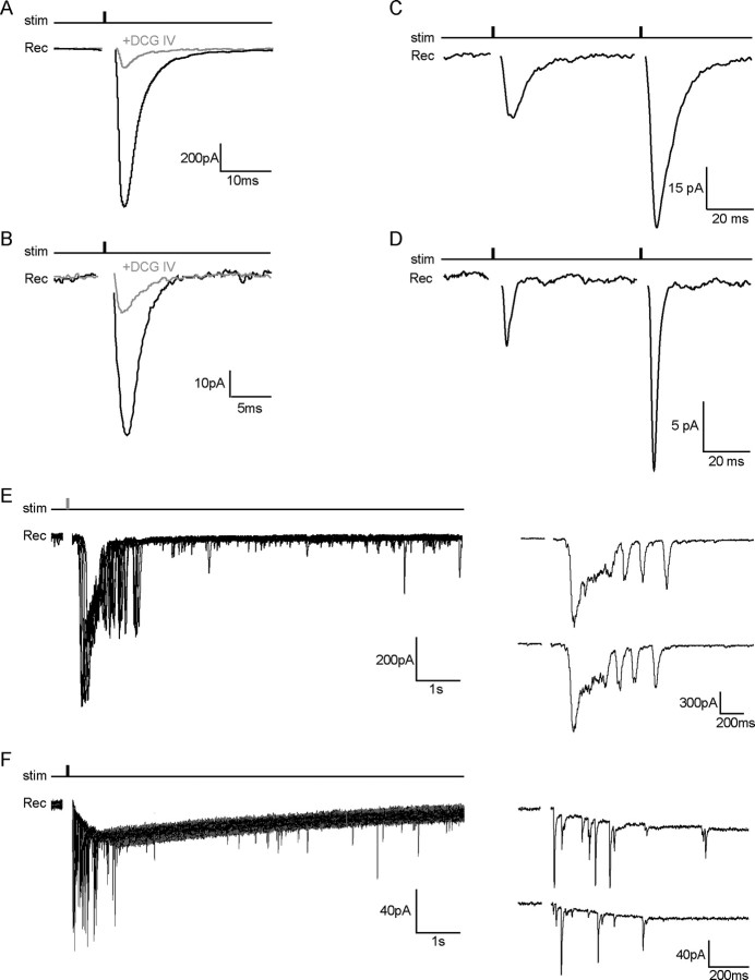
Figure 4.
sNG2+ cells and EGFP+ hilar interneurons receive glutamatergic inputs both from granule cells and CA3 pyramidal neurons. A, B, Evoked responses to granule cell layer stimulation in a hilar EGFP+ interneuron (A) (Vh = −60 mV) and its sNG2+ cell (B) (Vh = −80 mV) under control conditions (black trace) and in the presence of 1 μm DCG-IV (gray trace). Traces are averaged from 25 consecutive responses recorded from the same cell in the voltage-clamp mode (stimulus: 100 μs, 200 μA). The significant reduction of the evoked response after application of DCG-IV indicates that both cell types are contacted by mossy fiber synapses. Responses were evoked in an isolated dentate gyrus slice preparation. C, D, Facilitation of evoked responses by paired minimal stimulation of the granule cell layer (interstimulus interval, 50 ms) in a hilar EGFP+ interneuron (C) (Vh = −60 mV) and its sNG2+ cell (D) (Vh = −80 mV) (stimulus: 100 μs, 10 and 50 μA). The traces are averaged from 25 consecutive responses recorded from the same cell in the voltage-clamp mode. E, F, Superposition of consecutive responses evoked by CA3 layer stimulation in a hilar EGFP+ interneuron (E) (Vh = −60 mV) and its sNG2+ cell (F) (Vh = −80 mV) (stimulus: 100 μs, 200 μA). The right traces show examples of single responses evoked by CA3 layer stimulation in both cell types. All recordings were performed in the presence of 100 μm CTZ and 100 μm picrotoxin using a K-gluconate internal solution. stim, Stimulus; Rec, recording.

