
Fig. 1.
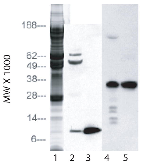
SDS/PAGE of concentrated stromal extract on 4–12% gels. Lane 1: Coomassie blue stain of 20 micro liters of extract. Lane 2: Western blot of 20 micro liters of extract using antibodies to IGF-II. Lane 3: Western blot of 2 micrograms of IGF-II using antibodies to IGF-II. Lane 4: Western blot of 20 micro liters of extract using antibodies to IGFBP. Lane 5: Western blot of 2 micrograms of IGFBP using antibodies to IGFBP.