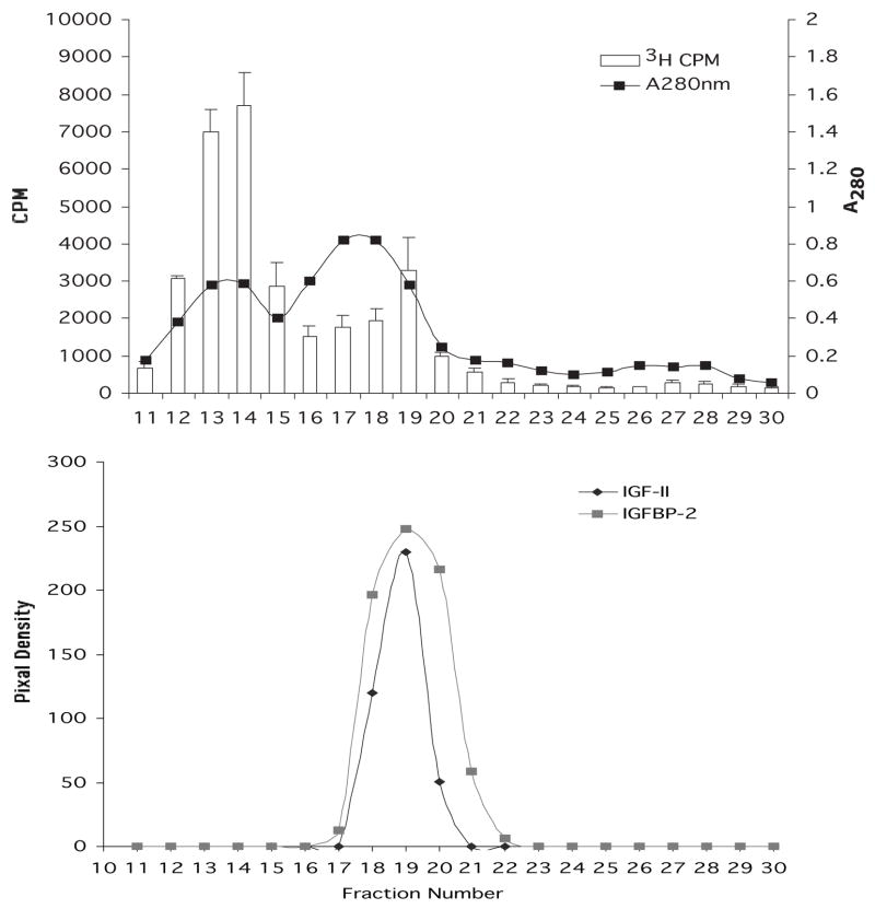
Fig. 2.
Chromatography of stromal extract on a column of Sephacryl S-300. Upper panel: fractions 11–30 were monitored for absorbance at 280 nm and for the ability to stimulate 3H thymidine incorporation in keratocytes in culture. Lower panel: fractions 11–30 were analyzed by Western blot using antibodies to IGF-II and to IGFBP. The pixel density of the IGF-II and IGFPB bands was measured and graphed.

