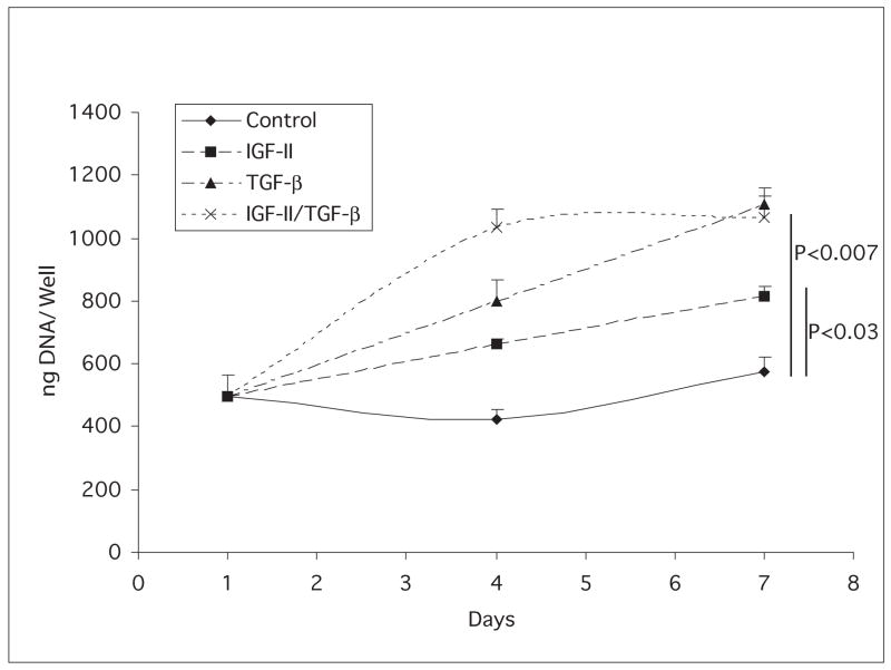
Fig. 3.
DNA content of keratocyte cultures. Keratocytes were cultured in media without (diamonds) or with IGF-II (squares), TGF-β (triangles) or IGF-II plus TGF-β (X). Compared to the DNA content of control day 1 cultures, the day 7 IGF-I, the day 7 TGF-β, and the day 7 IGF-II plus TGF-β cultures were significantly higher. P values are listed on figure.

