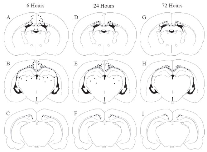
Figure 3. Anatomical and temporal distribution of axons exhibiting APP or RM014 immunoreactivity.
APP- (filled circles) and RM014-labeled (open circles) injured axonal profiles were located in the corpus callosum, cingulum, and lateral white matter tracts at 6 (A–C), 24 (D–F), and 72 hours (G–I) following diffuse brain injury. Note the greater extent of APP(+) immunoreactivity compared to RM014(+) axons in all brain regions.

