
Figure 1.
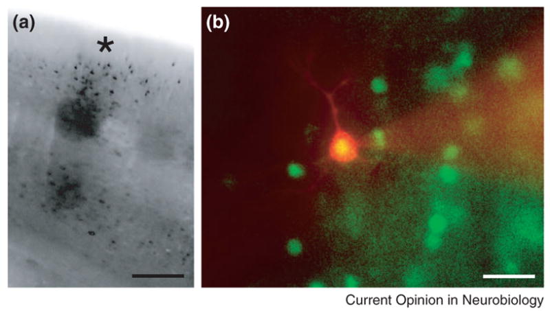
(A) FosGFP expression induced by in vivo sensory stimulation can be visualized in a single cortical column in vitro. Animals were subjected to removal of all but a single whisker from both sides of the mouse face for 24 hrs, a stimulus that results in robust fos expression. Cells expressing fosGFP can be detected in the single barrel column (*) representing this whisker in acute brain slices prepared from these animals. Figure modified from [6]. Scale ~150 μm. (B) A fosGFP+ Alexa-568 (red) filled cell after whole-cell recording. FosGFP+ neurons possess normal electrophysiological responses properties compared to neighboring, unlabeled cells [6]. Scale ~15 μm.