
Figure 2. Changes in AQP0 immunolabelling as a function of fiber cell differentiation.
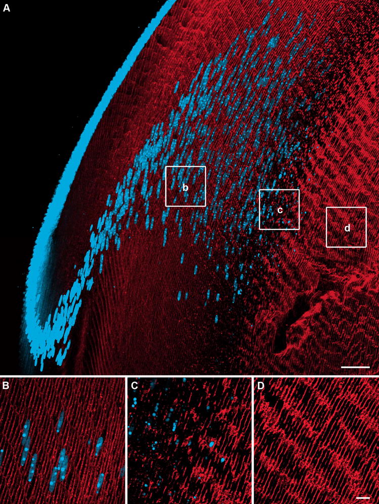
Axial cryosections of a rat lens double labelled with an AQP0 antibody (red) and the nuclear stain propidium iodide (blue). (A) Overview of AQP0 expression in the outer cortex, showing dispersal and degradation of cell nuclei. (B) In differentiating fiber cells AQP0 expression is predominantly membranous. (C) Just prior to nuclear degradation, a decrease in AQP0 labelling is seen. (D). Mature fiber cells in the inner cortex show a dramatic increase in AQP0 labelling following nuclei loss. Scale bars: A = 50 μm; B–D = 10 μm.