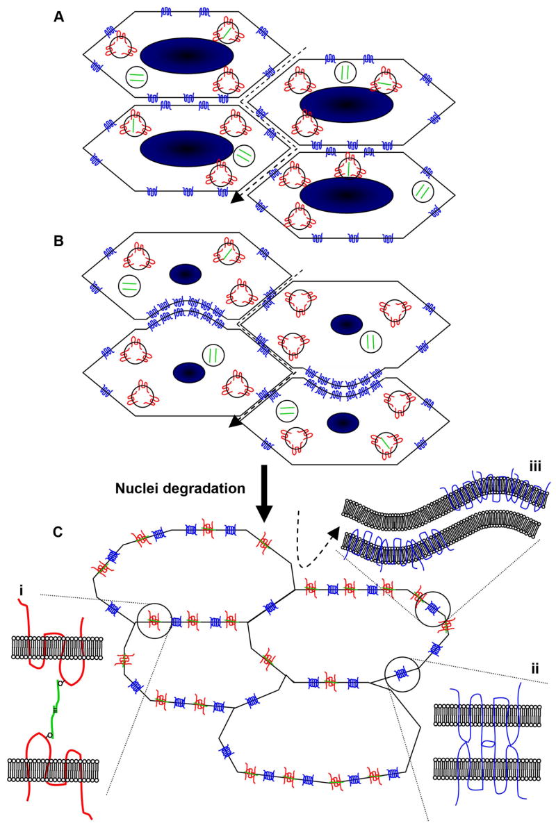
Figure 8. Model for interaction of MP20, galectin-3 and AQP0 in formation of the extracellular diffusion barrier.
(A) Nucleated cortical fiber cells contain intracellular vesicles of MP20 (red) and galectin-3 (green), while AQP0 (blue) is expressed predominantly in the cell membrane. The passage of large molecules (dashed arrow) through the extracellular space is possible. (B) Prior to degradation of cell nuclei, AQP0 redistributes in the membrane to broad side plaque-like structures, often in dome-like invaginations. (C) Upon nuclear degradation, MP20 and galectin-3 relocate to the cell membrane, and interact (i) to greatly attenuate the extracellular space. AQP0 molecules are brought into close apposition to form a junctional structure (ii). Alternatively, alternating aggregates of AQP0 in apposing cell membranes could lead to the formation of wavy junctions (iii). The passage of large molecules through the extracellular space is obliterated.

