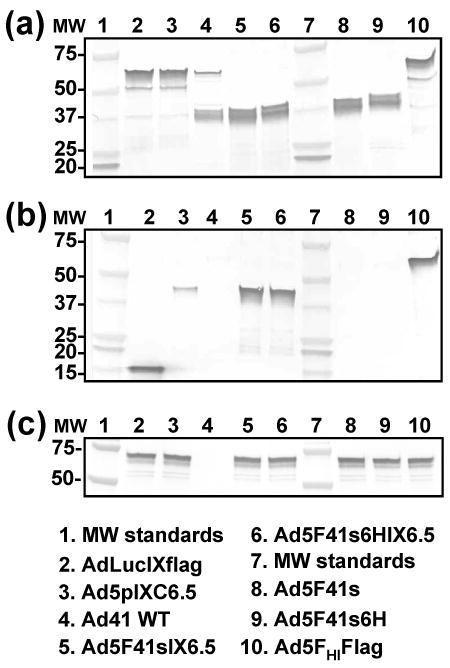
Fig. 4.

Western blot analysis of fiber and pIX modifications introduced into generated Ad vectors. Electrophoretically resolved viral proteins were transferred to a PVDF membrane and probed with mAb 4D2, mAb M2, or penton-base specific rabbit sera to detect the presence of fiber (a), pIX (b), or penton base (c), respectively, in the capsids of Ad5F41sIX6.5, Ad5F41s6HIX6.5, Ad5F41s, and Ad5F41s6H. AdLucIXflag, Ad5pIXC6.5, Ad41, and Ad5FHIFlag vectors served as controls. The numbers on the left indicate molecular masses of protein standards (lanes 1 and 7) in kDa.
