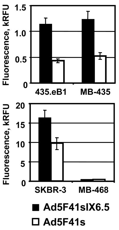
Fig. 5.
Evaluation of Ad5F41sIX6.5 infection via pIX-incorporated scFv. Monolayers of 435.eB1 and MDA-MB-435 cells (upper panel) or SKBR-3 and MDA-MB-468 cells (lower panel) were infected with Ad5F41sIX6.5 or control Ad5F41s vector at a dose of 100 vp/cell and incubated for two days to allow expression of the DsRed2 reporter gene. The fluorescent light intensity was measured in plate reader, using 560 nm emission and 620 nm excitation filters, and relative fluorescent units (RFU) detected in infected cells are presented as kRFU after subtracting the background light signal detected in uninfected cells. Each bar represents the cumulative mean of triplicate determinations ±SD.

