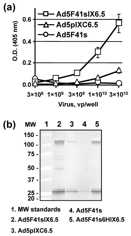
Fig. 6.

Validation of binding of Ad5F41sIX6.5-incorporated scFv to c-erbB2. (a) Ad5F41sIX6.5, Ad5pIXC6.5, and Ad5F41s viral particles were adsorbed on an ELISA plate at the indicated concentrations and incubated with 50-μl aliquots of SKBR-3 cell lysate. Bound c-erbB2 oncoprotein was detected with mAb cocktail against the cytoplasmic c-erbB2 domain followed by AP-conjugated goat anti-mouse secondary antibody, and the plate was read at 405 nm. Each data point represents the cumulative mean ±SD of triplicate determinations (some error bars are smaller than the symbols). (b) The indicated Ad vectors were incubated with ErbB2/Fc protein that was immobilized on protein A gel. The viral particles bound to ErbB2 were immunoprecipitated, eluted from the gel and analyzed by western blot using rabbit anti-Ad5 serum. The numbers on the left indicate molecular masses of protein standards in kDa.
