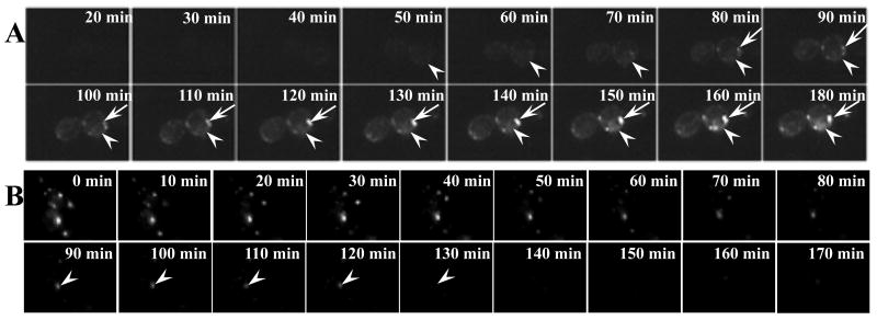
Fig. 3.
EGFP-CFTR localized within ERACs is degraded via a proteasome-independent pathway. (A) The proteasome-deficient pre1-1 yeast strain containing the EGFP-CFTR plasmid was imaged following induction of expression. The first image was captured ∼20 min after induction. Subsequent images were taken every 2 min. Movie montages at indicated time points are shown. CFTR in the pre1-1 strain forms ERACs (arrows). (B) The pre1-1 yeast strain was induced with copper for 2 h to form EGFP-CFTR foci followed by time-lapse imaging as described in Figure 1C. The arrowhead in (B) shows the rapid clearance of ERACs in the proteasome-deficient strain.

