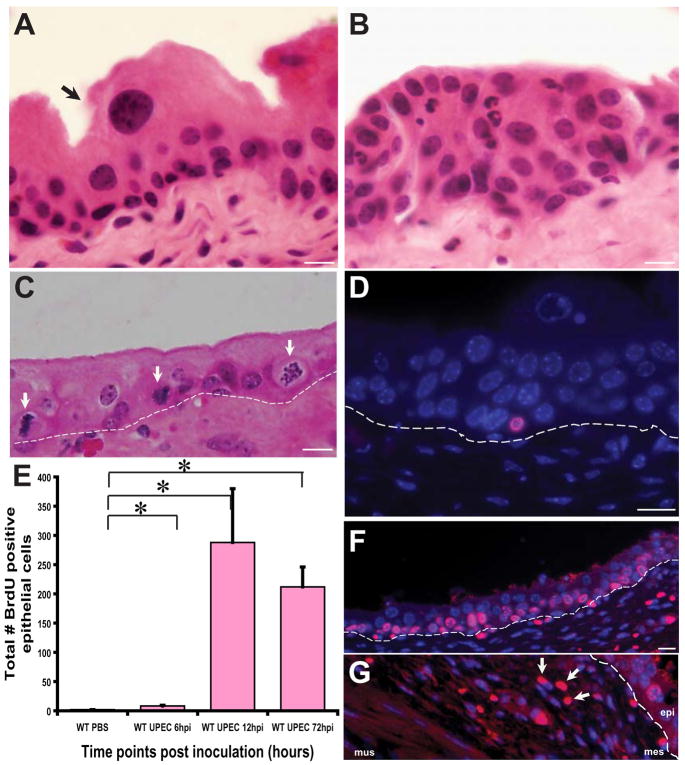
Figure 1. UPEC infection accelerates urothelial renewal.
(A) Hematoxylin and eosin (H&E) stained normal adult urothelium depicting an unperturbed USC niche. Arrows point to a mature superficial cell. (B) 12 hpi (hpi= hours post inoculation), there is exfoliation of mature superficial cells and hyperproliferation of the underlying immature basal cells concomitant with an inflammatory response. (C) Numerous mitotic figures (arrows) are evident localized to the basal layer housing presumptive stem cells. Dotted lines indicate the epithelial-mesenchymal boundary. (D) Immunofluorescence (IF) analysis reveals that BrdU (stained red with AlexaFluor 594-tagged anti-goat secondary antibodies) labels only basal cells at 6hpi. Nuclei are stained blue with biz-benzimide. (E) Total number of BrdU+ epithelial cells per tissue section (n= 1–2 sections/bladder; 4–7 mouse bladders/timepoint/condition) were counted. Depicted are data from 6h, 12h and 72h post UPEC inoculation relative to mock-infected bladders; p<0.05. P values were computed using a two-tailed Mann-Whitney test. (F) IF analysis of UPEC infected bladders at 72 hpi shows numerous BrdU+ cells in the USC niche. (G) BrdU+ mesenchymal cells are also evident (arrows) in the USC niche at 12 hpi and 72hpi. Bar= 10 μm.

