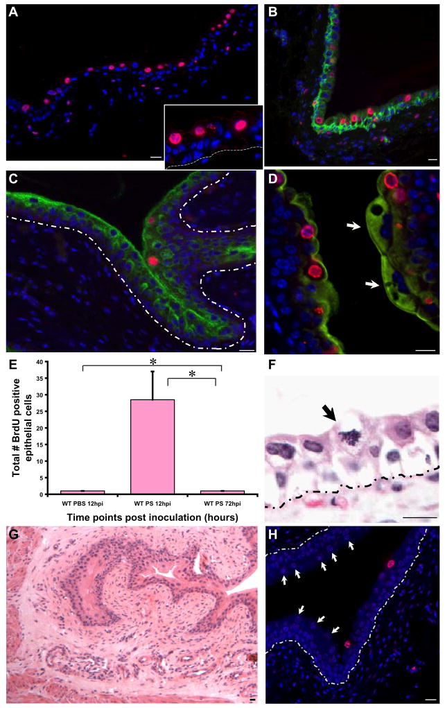
Figure 6. PS treatment activates transiently amplifying cell populations.
(A) IF analysis reveals that BrdU (stained red with AlexaFluor 594-tagged anti-goat secondary antibodies) labels non-basally located (intermediate) cells (shown in higher magnification in A_Inset). Nuclei are stained blue with biz-benzimide. Dotted lines indicate the epithelial-mesenchymal boundary. (B) BrdU+ cells (red) do not co-label with the cytokeratins 5, 6 (green)-expressing basal urothelial cell layer. (C) IF analysis of a 12h PS treated bladder, showing slight colocalization between BrdU (red) and uroplakin III, a terminal differentiation marker (stained green with AlexaFluor 488-tagged anti-mouse secondary antibodies). (D) Higher magnification, with a mature, multinucleated superficial facet cell densely expressing uroplakin III (arrow) opposite several BrdU+ intermediate cells. (E) Total number of BrdU+ epithelial cells were counted in 12h and 72h post PS treated bladders (n= 1–2 sections/bladder; 4–7 mouse bladders/timepoint/condition). (F) H&E stained PS treated bladders showing a mitotic figure in the suprabasal layer of the urothelium (arrow). (G) H & E stained 12h PS treated wild type bladder, showing no evidence of an inflammatory infiltrate. (H) IF analysis of BrdU+ cells in PS treated wild type bladders, PS treated for 72h, with few proliferating cells in suprabasal layer with presence of numerous regenerated mature facet cells present (arrows). Bar= 10 μm.

