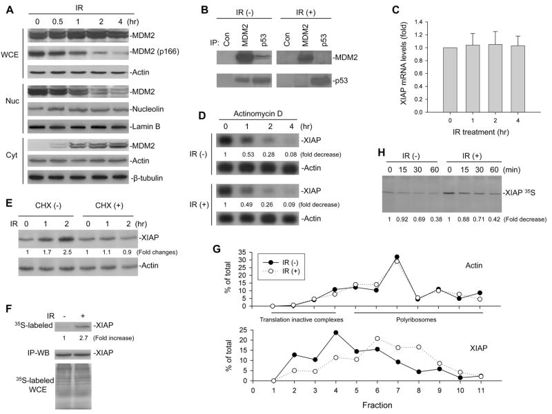
Figure 1.
Modulation of MDM2 and XIAP translation by IR. (A) EU-1 cells were treated with 10 Gy IR for different times as indicated, and cell extracts were tested for the nuclear (Nuc) and cytoplasmic (Cyt) expression of MDM2 as well as for total MDM2 and MDM2 phosphorylation at site 166 in whole cell extract (WCE) in western blot assays. (B) Cell lysates from EU-1 treated with or without 10 Gy IR for 4 hours were IP with antibodies as indicated. Normal mouse antibody served as control (Con). Proteins in immune complexes were detected by western blotting. (C) mRNA expression of XIAP after treatment with 10 Gy IR for the indicated time in EU-1 cells was determined by quantitative RT-PCR. Data represents the mean (±SD) levels of XIAP mRNA of three independent experiments. (D) EU-1 cells were treated with 10 Gy IR for 2 hours followed by addition of 5 mg/ml actinomycin D. At different times after actinomycin D addition, the cells were harvested, and total RNA was isolated. The amount of XIAP mRNA remaining was determined by Northern blotting and quantified by densitometric analysis. Labels under bands in the blot represent XIAP mRNA levels after normalization to actin, compared with samples (0, defined as 1 unit). (E) EU-1 cells were treated with and without 10μg/ml CHX for 10 min, before 10 Gy IR. Extracts from cells that were unirradiated (0) or harvested 1 or 2 h after IR were assessed for expression of XIAP, to show that CHX blocked IR-induced XIAP. (F) XIAP protein was IP from EU-1 cells that had been labeled for 5 min with [35S]-methionine, 30 min after 0 or 10 Gy IR, and then assessed by autoradiography (upper panel). Cells that had been pretreated with 50 μM MG132 and analyzed by western blot showed equivalent amounts of XIAP in the IP as a control for total XIAP protein that was not turnover in unirradiated versus irradiated cells (middle panel). Controls include analysis of whole cell extracts (WCE), where equal amounts of [35S]-methionine incorporated into the irradiated and unirradiated cells (bottom panel). (G) IR enhances association of the XIAP mRNA with translating polyribosomes. EU-1 cells were treated with or without 10 Gy IR for 12 h, and cytoplasmic lysates were fractionated on sucrose gradient. RNA was extracted from each of the fractions and subjected to quantitative RT-PCR for quantitative analysis of the distribution of XIAP and Actin mRNAs. Data represent percentage of the total amount of corresponding mRNA on each fraction. (H) Turnover of XIAP protein in IR-treated and control cells, as detected by pulse-chase assay. EU-1 cells were treated with or without 10 Gy IR for 2 hours followed by [35S]methionine labeling. At selected times as indicated after labeling cell lysates were collected for determination of the XIAP 35S protein levels by SDS-PAGE analysis.

