Figure 2.
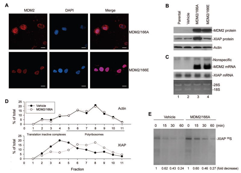
Effect of cytoplasmic MDM2 on XIAP transcription, translation, and post-translational modification. (A) Subcellular distribution of transfected MDM2/166A and MDM2/166E, as demonstrated by the red fluorophores in EU-4 cells characterized by confocal microscopy. Scale bars = 10μm. (B) and (C) Expression of transfected MDM2 and XIAP in both MDM2-transfected and control cells were detected by western blot assay (B), and their mRNA levels were detected by Northern blot assay (C). (D) Quantitative RT-PCR analysis of the MDM2/166A-induced translation enhancement of XIAP mRNA. SH-SY5Y cells were transfected with MDM2/166A and control plasmid (Vehicle), and cytoplasmic extracts were subjected to linear sucrose gradient fractionation. RNA was extracted from each of the fractions and quantitatively analyzed to evaluate polyribosome association by the XIAP and Actin mRNA. (E) Turnover of XIAP protein in MDM2/166A-transfected and control cells, as detected by pulse-chase assay. The radioactive labeled XIAP protein levels were quantified by densitometric analysis. All values were corrected relative to the load control and labeled under bands in the blot compared with samples (0, defined as 1 unit).

