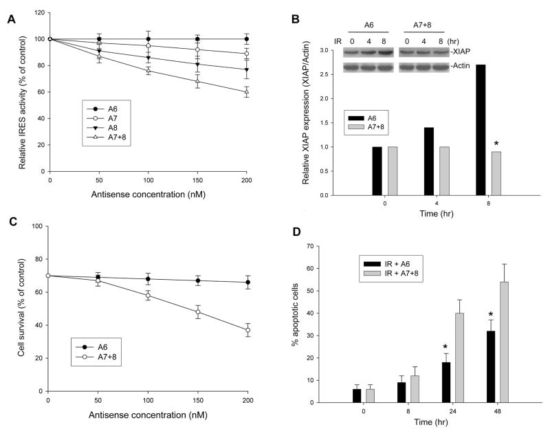
Figure 7.
Effect of blockage between MDM2 and XIAP IRES interaction on XIAP expression and IR-induced apoptosis. (A) LA1-5S cells were co-transfected with 5μg pβgal-xi-CAT plasmid and increasing amounts of antisense (A6, A7, A8 and A7+8 as shown in Fig. 5C). The % of CAT activity (mean ± SD, calculated by normalizing with βgal activity) was given relative to a translation reaction performed in the absence of antisense. (B) The effect of antisense (A6 and A7+8) on IR-induced upregulation of endogenous XIAP in LA1-5S cells. Cells transfected with 200 nM A6 or A7+8 were exposed to IR for the indicated time. The expression levels of XIAP, using Actin as a control, were analyzed by western blotting (insert). Data in the graph represent the relative expression of XIAP as compared to Actin after this western blot was densitometrically scanned. *p<0.01. (C) LA1-5S cells were treated with 10 Gy IR in the presence or absence of different dose of A6 or A7+8 as indicated. Cells were incubated for 48 h, and cell viability was determined by WST assay. Data represent the mean percentage (±SD) of cell survival from three independent experiments. (D) Time-course of apoptosis induced by IR in LA1-5S cells in the presence of A6 or A7+8 antisense (200 nM). Cells were treated with 10 Gy IR for the indicated time points, and apoptotic cells were detected by annexin-V staining using flow cytometry. Data represent the mean percentage of annexin-V positive cells from three independent experiments; bars, ± SD. *p<0.01.

