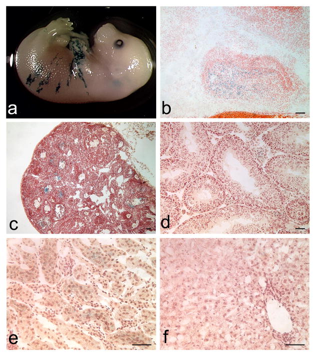
Figure 3. Minimal ectopic Tg(Gh-cre)SAC1 transgene expression.
Very limited X-gal staining is detected in non-pituitary tissues such as the skin surrounding the limbs and trunk of developing embryos (a). At e18.5, X-gal staining is appropriately restricted to the developing anterior lobe (A) and is absent from the intermediate (I) and posterior (P) lobes (b). In adult tissues, X-gal staining is seen in the developing follicles of some ovaries (c). Sporadic but very faint X-gal staining appears in the Leydig cells of the testes (d) and in the renal cortex (e). No staining is detected in the adult liver (f). All magnification bars represent 50μm.

