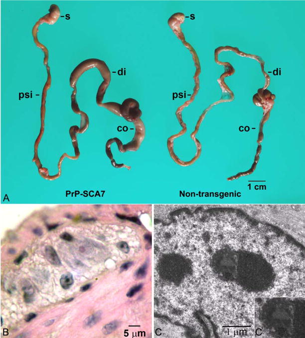
Figure 1.
(A) A photograph of the gastrointestinal tracts from a symptomatic, 15-week, PrP-SCA7-92Q mouse and its non-transgenic littermate demonstrates marked distension of the proximal colon (co) and distal small intestine (di) of the transgenic animal. The stomach (s) and proximal small intestine (psi) are not affected. (B) A photomicrograph from an H&E-stained section demonstrates a rare eosinophilic intranuclear inclusion (arrowhead) in a myenteric neuron from a transgenic mouse. (C) Ultrastructurally, a subset of neurons in transgenic mice contained granular electron dense inclusions (arrowhead and inset), which appeared more heterogenous than neighboring nucleoli (n).

