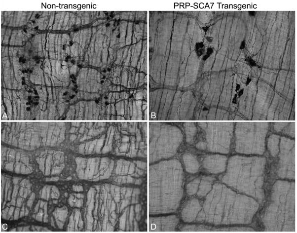
Figure 6.
(A, B) Wholemount preparations of distal ileum from non-transgenic (A) and PrP-SCA7-92Q transgenic, 15-week-old mice (B) mice stained histochemically for NADPH-diaphorase (nNOS) activity demonstrate reduction in the density of nNOS-cell bodies in the transgenic animals. Those nNOS neurons that remain are enlarged, but the network of nNOS-positive nerve fibers is simplified with fewer and thinner cell processes. (C, D) Acetylcholinesterase histochemical staining of similar preparations also demonstrates reduction of the density and number of nerve processes in the myenteric plexus of a PrP-SCA7-92Q transgenic mouse (D) compared to its non-transgenic littermate (C).

