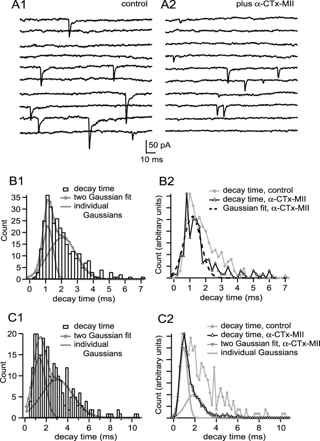
Figure 6.
mEPSC populations with a rapid decay time, characteristic of pharmacologically isolated α7-nAChR mEPSCs, are common. A1 shows examples of mEPSCs recorded from a cell having fast rising mEPSCs with variable decay (10 sequential responses). B1 shows a decay time probability density function of 297 events; the function was best fit by the sum of two Gaussians, with modes of 1.17 and 2.13 ms. Addition of α-CTx MII to block α3-nAChRs (examples in A2; 10 sequential responses) converted the probability density function into one best fit by a single Gaussian with a mode of 1.26 ms (B2). C1 shows a decay time probability distribution function for a different cell that was fit with two Gaussians having modes of 1.32 and 2.98 ms. Addition of α-CTx MII to this cell yielded, again, a two Gaussian population, with modes of 0.95 and 1.95 ms (C2). The mode of the fast Gaussian was not altered by α-CTx-MII, suggesting that the fast-decaying events are produced by activation solely of α7-nAChRs. See legend to Figure 3 for additional information regarding binning of decay time values.

