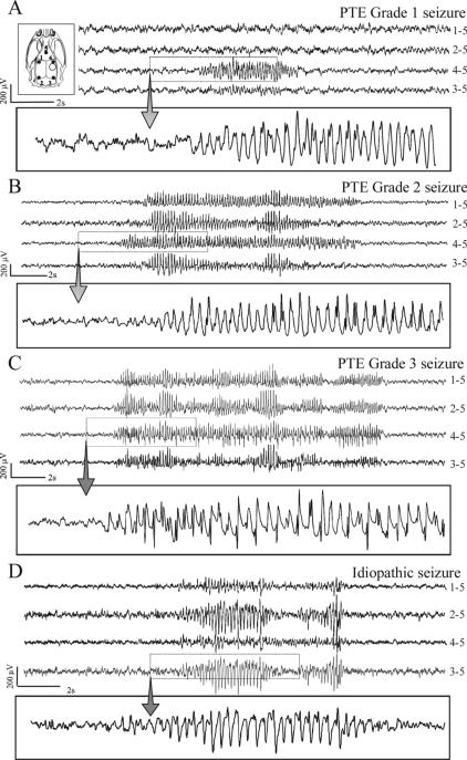
Figure 1. Different types of chronic recurrent seizures as revealed by ECoG following FPI.
Inset top, left) Schematic of the locations of the five cortical electrodes (filled circles) and of the injury site (hollow circle) in respect to the rat skull. All panels represent continuous ECoG monitoring at 7 months post-FPI in wake animals. A) A representative grade 1 posttraumatic seizure detected exclusively by the peri-lesion electrode. B) A representative grade 2 posttraumatic seizure first detected by the peri-lesion electrode and then by multiple channels. C) A representative grade 3 posttraumatic seizure detected simultaneously by multiple channels. D) A representative idiopathic seizure, detected bilaterally and characterized by larger occipital epileptic discharge. These idiopathic seizures were absent until 5.5 months of age and represented 3.6% of all cortical discharge of FPI animals at 8 months of age. ECoG calibration bars are on the left of each panel. Dotted boxes highlight the portion of ECoG shown at higher temporal resolution in the rectangle underneath each panel. The numbers next to each ECoG trace indicate the electrodes by which the trace is recorded, and its reference.

