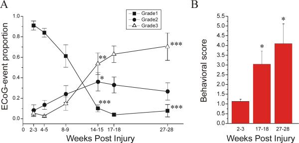
Figure 3. Temporal evolution of the FPI-induced posttraumatic epileptic syndrome.
Electrical and behavioral correlates of PTE progression are assessed in 8 epileptic animals. A) The proportions of ECoG events of grade 1, 2, and 3 are plotted over time from 2 to 28 weeks post-injury. Focal frontal-parietal seizures (grade 1; filled square) represented the most common seizure type 2−3 weeks post-injury, but then progressively decreased in proportion over time. Focal neocortical spreading seizures (grade 2; filled circles) were rare at 2−3 weeks post-injury, but then increased in proportion over time and peaked at 14−15 weeks post-injury. Seizures that did not originate from the frontal-parietal cortex and appeared bilateral in onset (grade 3; hollow triangle) were rare up to 8−9 weeks post-injury, and then increased up to 27−28 weeks post-injury. Note the overall increase in seizures’ bilateral spread over time post-injury (grades 2&3 combined). B) The behavioral score during epileptiform ECoG events is plotted versus time post-injury. Note the progression of the severity of behavioral seizures over time post-injury. Data are presented as Mean±S.E.M. Statistics with paired t-test (* p<0.05; ** P≤0.01; *** P≤0.001, vs 2−3 weeks).

