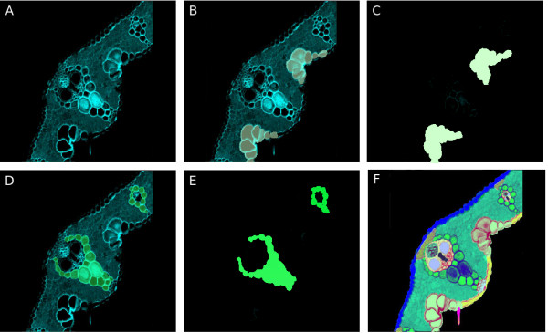
Figure 3.

Creation of a map for the transversal section of a rice leaf. A. Confocal Image of a rice leaf section. B. Using image processing software, the leaf section image is divided into two layers, and the region occupied by bulliform cells is colored. C. The map of bulliform cells for the leaf section is obtained by saving the colored layer as a different archive. D, E. By repeating the process described previously in B and C, the map for the bundle sheath region is obtained. F. By overlaying all obtained maps for all regions identified in the leaf section, we can construct the full map for the picture, and when overlaid with the original picture, each region becomes distinctively painted in a different color.
