
Figure 3.
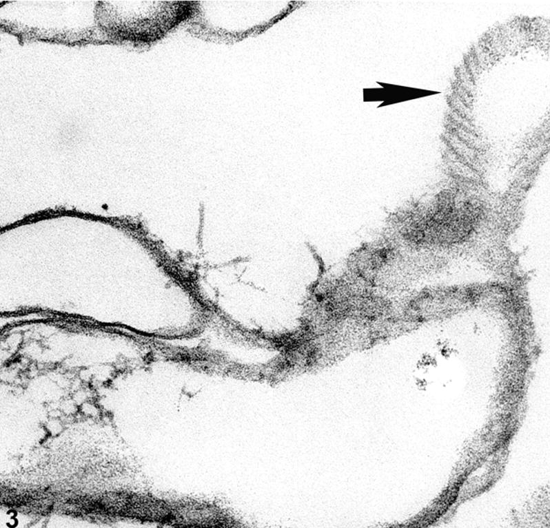
Electron micrograph of ghosted mouse lens, showing a tangential view of a fiber cell membrane (arrow) where IFs are aligned along the surface of the membrane.
Official websites use .gov
A
.gov website belongs to an official
government organization in the United States.
Secure .gov websites use HTTPS
A lock (
) or https:// means you've safely
connected to the .gov website. Share sensitive
information only on official, secure websites.
Electron micrograph of ghosted mouse lens, showing a tangential view of a fiber cell membrane (arrow) where IFs are aligned along the surface of the membrane.