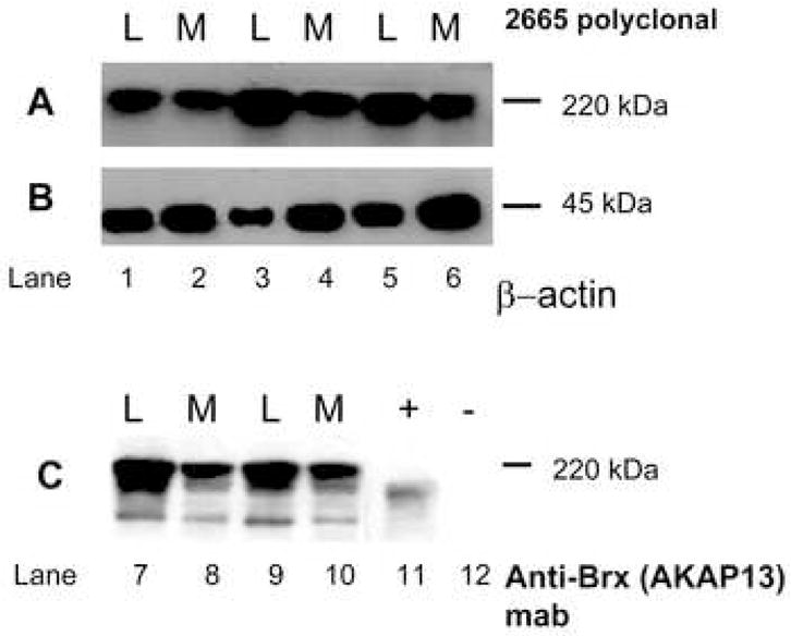
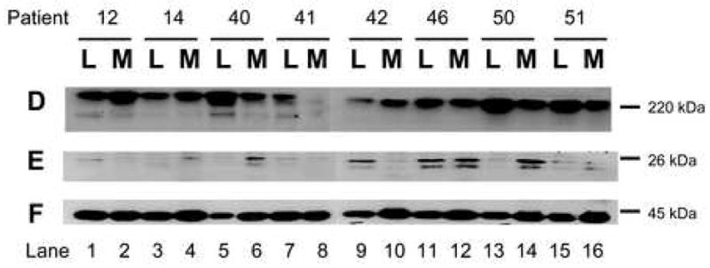
Figure 3. Altered expression of factors involved in mechanical transduction in matched surgical specimens of uterine leiomyoma and myometrium.


3A: Western analysis of leiomyoma (L) and myometrial (M) lysates for expression of the Rho-GEF AKAP13 using 2665 affinity-purified antisera.29 A 220kDa band was present that was increased in leiomyoma compared to myometrium. Samples from patients 12–16, 40–41, 50, 51 were used for all western blots.
3B: Beta actin control for lysates in A.
3C: Western analysis for AKAP13 using monoclonal antibody directed against Brx (Upstate, Temecula, CA). A 220kDa band is identified. Positive control (+) consisted of lysates prepared from Cos-7 cells transfected with a construct expressing a 170kDa form of AKAP13. The negative control (−), lane 12, were lysates prepared from un-transfected Cos-7 cells (Cos-7 cells do not express AKAP13).
3D: Western analysis for AKAP13 in matched leiomyoma (L) and myometrial (M) lysates from 8 patients. In most, but not all pairs, expression of AKAP13 was greater in leiomyoma (L) compared to myometrium (M).
3E: Western analysis for RhoA in the same lysates. Readily extractable levels of RhoA were increased in some, but not all fibroid samples.
3F: Beta actin control for protein loading.
